
FIG. 1.
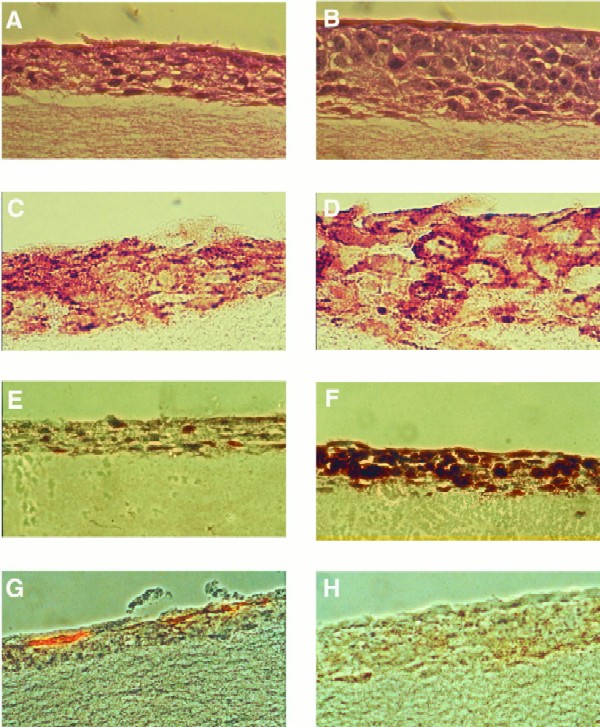
Characterization of HaCaT cell organotypic raft cultures. Hematoxylin-eosin stains of vector control (A) and LMP2A-expressing (B) HaCaT rafts are shown. LMP2A rafts are thickened with rounded cells containing large nuclei. Cells in vector control rafts are flattened, and enucleated cells are evident in the top layers of the culture. LMP2A expression was detected in the plasma membrane of cells in all layers of the epithelium using a rabbit HA antiserum and a biotin-streptavidin-peroxidase detection system (DAKO) (D). Background staining is evident in vector control rafts (C). Cell proliferation was determined by BrdU incorporation and staining with an anti-BrdU monoclonal antibody. Single BrdU-positive cells were observed in vector control rafts (E), while LMP2A-expressing rafts were highly proliferative (F). Localization of proliferating cells was not restricted to the basal cell layer. LMP2A blocks cell differentiation. The differentiation marker involucrin was present in the topmost layers of the epithelium of vector control rafts (G) but was not detected in LMP2A-expressing rafts (H).