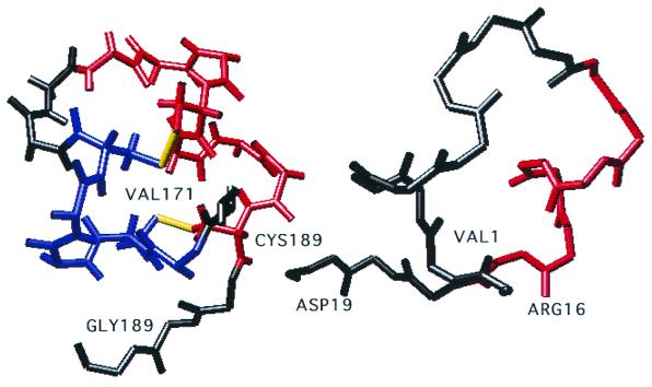
FIG. 6.
Representation of the superimposition of the cysteine noose of BRSV strain 391.2 (left) and of the consensus sequence of isolates from subgroup VI (right). α Helices are indicated in blue and red. Disulfide bridges are drawn in yellow. Residues 171 to 189 in the representation of strain 391.2 correspond to residues 1 to 19 in that of subgroup VI.

