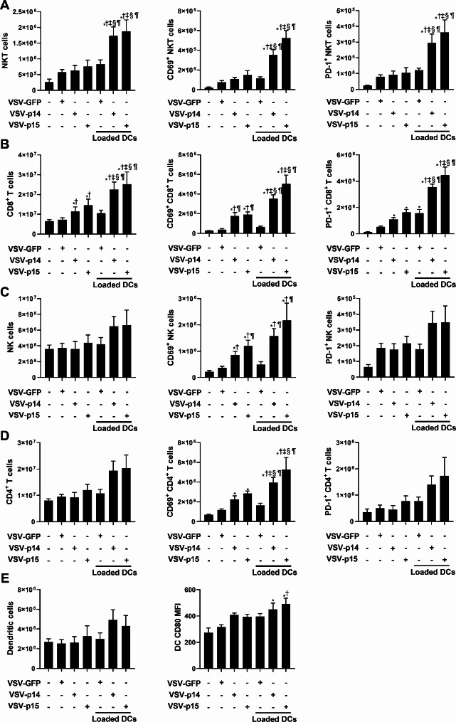
Fig. 7.
VSV-p14 or VSV-p15 in combination with NKT cell activation increases immune activation. VSV and NKT cell immunotherapy treatments were administered in the metastatic 4T1 model as outlined in Fig. 5A. Spleens from untreated and treated mice were isolated seven days after treatment. Flow cytometry was used to assess immune cell expansion and activation (n = 5–9 per group). The number of (A) NKT cells (CD1d tetramer+ TCRβ+), (B) NK cells (NK1.1+ TCRβ−) (C) CD8+ T cells (TCRβ+ CD8α+), (D) CD4+ T cells (TCRβ+ CD4+) and the expression of CD69, PD-1, and intracellular IFNγ by these subsets was assessed. (E) The number of dendritic cells (MHC II+ CD11c+) and CD80 expression were also examined. *p < 0.05 compared to untreated, †p < 0.05 compared to VSV-GFP, ‡p < 0.05 compared to VSV-p14, §p < 0.05 compared to VSV-p15, ¶p < 0.05 compared to VSV-GFP + glycolipid-loaded DCs

