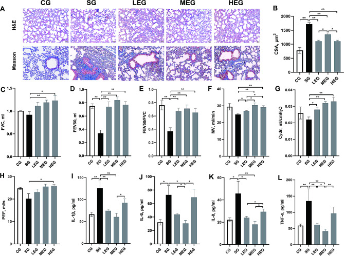
Fig. 1.
Features of COPD changes after exercise training. A, lung sections stained with HE and Masson (Bar = 50 μm, 100× and 200×, respectively); B, CSA of alveolar; C-H, parameters of lung volume and ventilation function; I-L, cytokines level of BALF. CSA, cross sectional area; FVC, forced vital capacity; FEV50, forced expiratory volume in 50 ms; MV, minute ventilation; Cydn, dynamic lung compliance; PEF, peak expiratory flow; IL, Interleukin; TNF, tumor necrosis factor; CG, control group; SG, cigarette smoke group; LEG, low-intensity exercise group; MEG, moderate-intensity exercise group; HEG, high-intensity exercise group; *p < 0.05; **p < 0.01

