Figure 1.

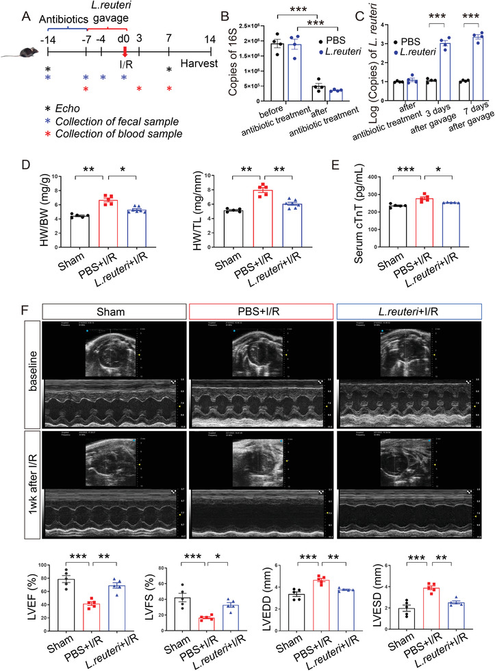
Pretreatment of L. reuteri ameliorated cardiac damage after I/R. A) Schematics of the experiment with L. reuteri gavage. Male C57BL/6J mice were pretreated with 7 days of antibiotics to remove gut microbiota and 7 days of L. reuteri gavage before I/R. Serum samples were collected at 7 days before I/R, 3 days, and 7 days after I/R. Fecal samples were collected before and after antibiotic treatment, 3 days and 7 days after L. reuteri gavage. Cardiac function was detected by transthoracic echocardiography (Echo) at baseline and 7 days after I/R. Heart tissues were harvested at 14 days after I/R for histological assessment. B) The copies of bacterial 16S detected in the feces collected before and after antibiotic treatment. Data are shown as the mean ± SEMs, n = 4 per group. ***, p < 0.001 (Student's t‐test). C) The copies of L. reuteri detected in the feces collected after antibiotic treatment and at 3 days and 7 days after L. reuteri gavage. Data are shown as the mean ± SEMs, n = 4 per group. ***, p < 0.001 (Student's t‐test). D) Ratio of heart weight to body weight (HW/BW) and heart weight to tibial length (HW/TL) at 2 weeks after I/R. Data are shown as the mean ± SEMs. Sham group (Sham), n = 5; PBS‐gavaged I/R group (PBS+I/R), n = 5; L. reuteri‐gavaged I/R group (L. reuteri+I/R), n = 7. *, p < 0.05; **, p < 0.01 (one‐way ANOVA with post hoc Dunnett's test). E) Serum cTnT levels measured by ELISA at 3 days after I/R. n = 5 per group. *, p < 0.05; ***, p < 0.001 (one‐way ANOVA with post hoc Tukey test). (F) Representative echocardiographic images at baseline and 1 week after I/R. Quantitative data on left ventricular ejection fraction (LVEF), fractional shortening (LVFS), left ventricular end‐diastolic dimension (LVEDD), and left ventricular end‐systolic dimension (LVESD) at seven days after I/R are shown as the mean ± SEMs; n = 5 per group. *, p < 0.05; **, p < 0.01; ***, p < 0.001 (one‐way ANOVA with post hoc Tukey test). L. reuteri, Lactobacillus reuteri; I/R, ischemia/reperfusion. cTnT, cardiac troponin T.
