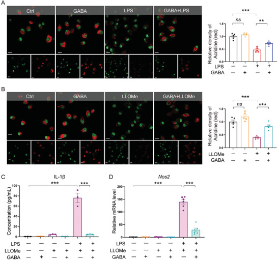
Figure 7.

GABA suppressed NLRP3 inflammasome activation by inhibiting macrophage lysosomal leakage. A) Acridine orange staining of BMDM under LPS stress showing lysosomal leakage. As a cell‐permeable green fluorophore that can be protonated and trapped in acidic vesicular organelles, acridine orange fluoresces red in intact lysosomes and fluoresces green elsewhere that is less acidic. Quantitative data shown as the mean ± SEMs to the right. n = 5 per group. ns, not significant; **, p < 0.01; ***, p < 0.001 (one‐way ANOVA with post hoc Tukey test). Scale bar, 10 µm. B) Acridine orange staining of BMDM treated with LLOMe or PBS in the presence or absence of GABA. Red fluorescence indicates intact lysosomes. Quantitative data are shown as the mean ± SEMs to the right. n = 5 per group. ns, not significant; ***, p < 0.001 (one‐way ANOVA with post hoc Tukey test). Scale bar, 10 µm. C) Measurement of IL‐1β secretion in cell culture supernatants by ELISA. Data are shown as the mean ± SEMs, n = 3 per group. ***, p < 0.001 (one‐way ANOVA with post hoc Tukey test). D) qRT‐PCR analysis of the mRNA levels of Nos2. n = 6 per group. ***, p < 0.001 (one‐way ANOVA with post hoc Tukey test). GABA, γ‐aminobutyric acid; LPS, Lipopolysaccharide; Nos2, Inducible nitric oxide synthase; IL‐1β, Interleukin‐1β.
