Figure 6.

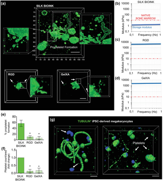
The unique softness of 3D bioprinted silk‐bioink is crucial to support thrombopoiesis. a), The silk bioink recreates a favorable environment for supporting increased proplatelet branching (top) compared to other conventional bioinks (bottom) (green = CD61; scale bar = 20 µm, representative of three independent experiments). b–d), Storage modulus of silk bioink compared to other conventional crosslinked bioinks b), silk bioink, c), RGD; d), GelXA). The red line identifies the maximal stiffness measured for the native bone marrow tissue. e), Percentage of proplatelet formation in the different tested conditions (n = 10; * p<0.001). f), Fold increase of platelet count/area in the different tested conditions (n = 10; * p<0.001). g), Human iPSC‐derived megakaryocytes (imMKCL) engineered to express fluorescent β1‐tubulin (green) show that proplatelet formation and platelet release are sustained by cytoskeleton remodeling (scale bar = 10 µm).
