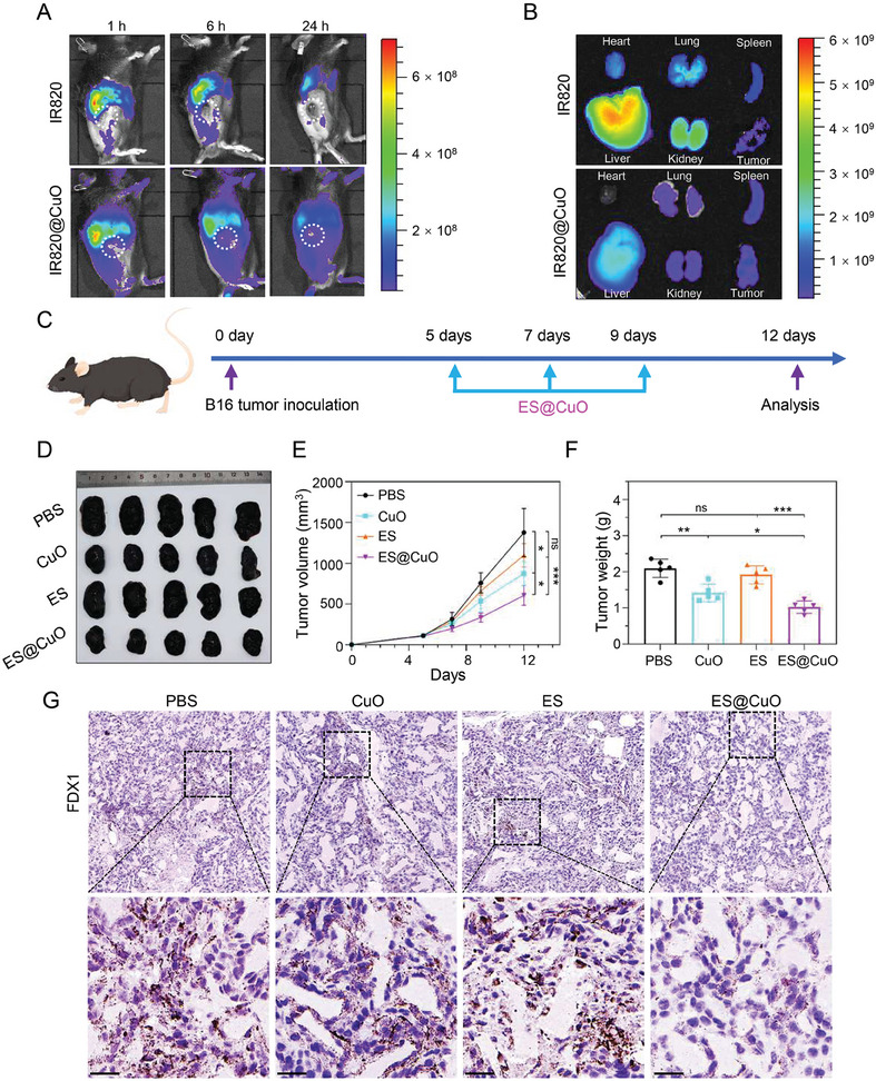
Figure 4.

Antitumor effects mediated by ES@CuO in vivo. A) Biodistribution after intravenous injection of free IR820 and IR820@CuO NPs at the indicated time points. B) Ex vivo imaging of IR820 fluorescence intensity in major organs and tumors collected at 6 h post‐injection. C) Schematic illustration of the experimental schedule for B16 tumor‐bearing mice. D) Corresponding tumor photographs, E) tumor growth curves, and F) ex vivo tumor weights of the B16 tumor‐bearing mice after various treatments (n = 5). G) IHC analysis of FDX1 expression in tumor tissues after different treatments. Scale bar: 25 µm. Data are shown as the mean ± SD; p values were calculated using an unpaired, 2‐tailed Student's t test with Welch's correction; ns, not significant; *p < 0.05; **p < 0.01; ***p < 0.001.
