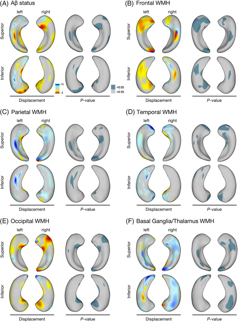
FIGURE 1.

Hippocampal shape alterations associated with Aβ status and regional WMH volumes in MITNEC‐C6 subjects. Regional hippocampal surface deformities associated with (A) Aβ positivity, and regional WMH load, localized to the (B) frontal lobe, (C) parietal lobe, (D) temporal lobe, (E) occipital lobe, and (F) basal ganglia/thalamus. The left side of each panel shows the relative displacement map (blue = outward displacement, red = inward displacement) associated with each disease process and the right side of each panel indicates the P‐value map related to each hippocampal surface. Aβ, amyloid beta; MITNEC‐C6, Medical Imaging Trials Network of Canada Project C6; WMH, white matter hyperintensities.
