
FIGURE 2.
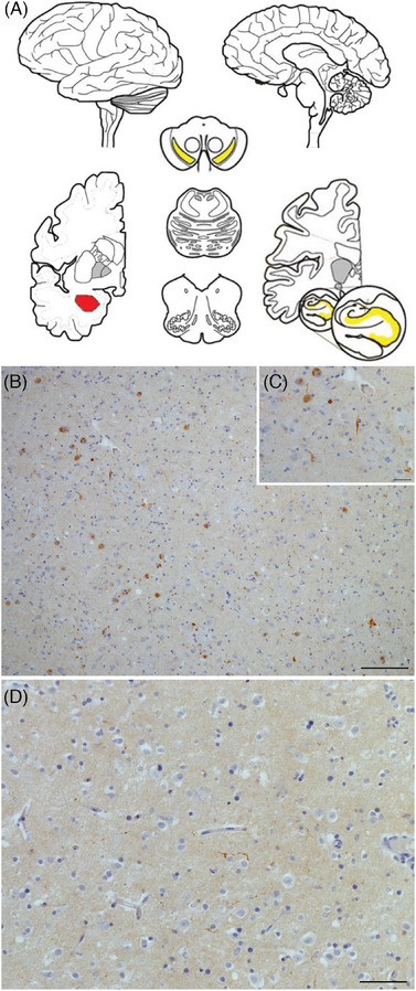
CSF αSyn‐SAA positivity is associated with spread in amygdala‐predominant LBD. (A) Schematic showing amygdala‐predominant LBD concentrated in amygdala (red); it may be confined there, or it may be found to a lesser degree in mesial temporal lobe structures (hippocampus or entorhinal cortex) or substantia nigra (yellow), adapted from Attems et al. (2021). 31 Immunohistochemical staining for all αSyn species in a representative case shows abundant Lewy bodies and Lewy neurites in the amygdala at low (10×, B) and high (40×, C) magnification with little LBD in the entorhinal cortex (20×, D). Scale bars: 100 μm (B), 25 μm (C), and 50 μm (D).