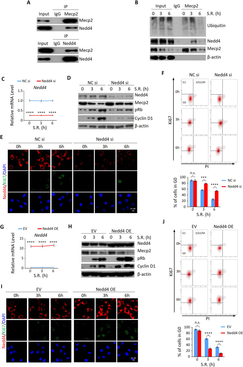
Figure 5. Nedd4 interacts with Mecp2 and affects quiescence exit by facilitating Mecp2 degradation.
(A) Reciprocal immunoprecipitation-western blotting (IP-WB) analysis to validate the interaction between endogenous Mecp2 and Nedd4. (B) Co-IP of Mecp2, ubiquitin, and Nedd4 in quiescent 3T3 cells during serum restimulation (SR)-induced quiescence exit. (C) Real-time PCR showing siRNA-mediated Nedd4 knockdown (KD) in 3T3 cells upon SR-induced quiescence exit. Data are presented as means ± SEM; n = 5. ****p<0.0001 by two-way ANOVA. (D–F) The effect of Nedd4 KD on quiescent exit in 3T3 cells determined by WB (D), immunofluorescence (IF) staining of Ki67 and Nedd4 (E), and Ki67/PI staining followed by flow cytometry (F) at the indicated time points. Lower panel in (F): quantification of the percentage of 3T3 cells in the G0 phase. Data are presented as means ± SEM; n = 3. n.s., not significant; ***p<0.001, ****p<0.0001 by two-way ANOVA. (G) Real-time PCR showing Nedd4 overexpression (OE) in 3T3 cells upon SR-induced quiescence exit. Data are presented as means ± SEM; n = 5. ****p<0.0001 by two-way ANOVA. (H–J) The effect of Nedd4 OE on quiescent exit in 3T3 cells determined by WB (H), IF staining of Ki67 and Nedd4 (I), and Ki67/PI staining (J) at the indicated time points. Data are presented as means ± SEM; n = 3. n.s., not significant; ****p<0.0001 by two-way ANOVA.


