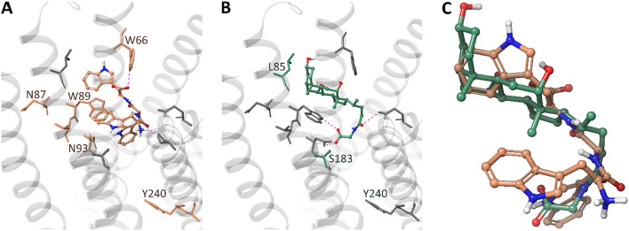
Fig. 8.
Putative binding modes of l-Trp-Trp-Trp (A) and glycocholic acid (B) into the TAS2R14 binding site. A zoom-on on the structural alignment of the docking poses is reported in panel C. Glycocholic acid and l-Trp-Trp-Trp structures are shown as CPK ball&stick with carbons in light orange and green, respectively. Binding site residues analysed by mutagenesis are shown as sticks. l-Trp-Trp-Trp specific residues are colored in light orange and GCA specific residues in green. Hydrogen bonds are displayed as magenta dashed lines

