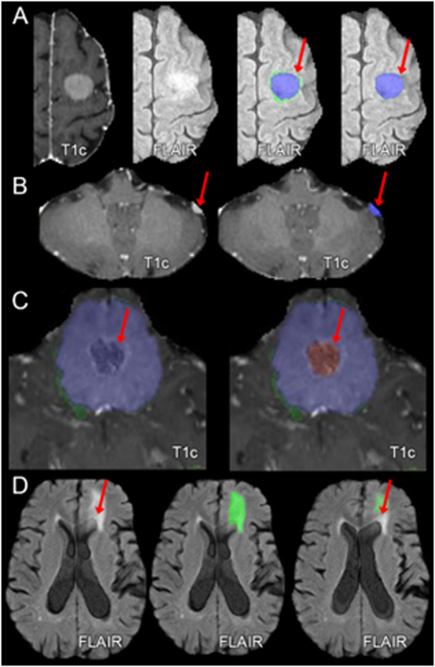
Fig. 3.

Examples of common errors of automated meningioma segmentation. (A) Erroneously marked a thin rim of edema that does not exist; (B) Missed small convexity meningioma; (C) Improper classification of non-enhancing tumor; (D) Tumor-related edema adjacent to presumed microvascular ischemic periventricular white matter FLAIR abnormalities. T1c: T1-weighted post-contrast imaging; FLAIR: T2-weighted fluid attenuated inversion recovery imaging.
