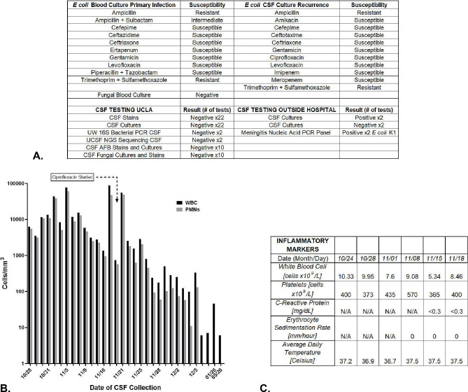
Fig. 1.
Cerebral Spinal Fluid (CSF), Serum, and Clinical Markers of Inflammation. A: Culture and susceptibility testing results from blood and CSF samples from the outside hospitals from the first and recurrent episode of E coli infection. Culture and testing results from UCLA after transfer. B: This graph demonstrates the pleocytosis measured in the CSF over the course of antibiotic treatment during the patient’s admission. Black bars demonstrate the levels of WBCs detected on sampling and the grey bars represent the proportion of those WBCs consisting of neutrophils. The start of ciprofloxacin is shown with a text box and arrow at the top of the graph. Neutrophil predominant CSF pleocytosis resolved after the introduction of ciprofloxacin. C: Serum and core temperature measurements of the patient at the time frame concerning for continued infection despite negative CSF gram stain, cultures, bacterial 16 S PCR testing, and unbiased pathogen NGS testing. All testing was negative for signs of systemic inflammation

