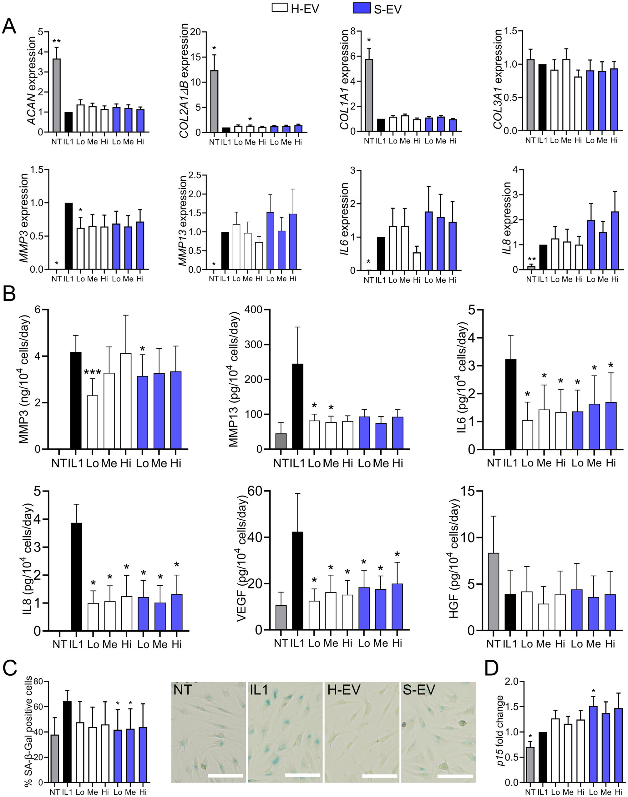
Fig. 3.

Senescent ASC-EVs exert a chondroprotective effect on OA chondrocytes cultured in resting conditions. Primary human chondrocytes were pretreated with 10 ng/mL IL1β (IL1) or not (NT) for 48 h. Different amounts (Low (Lo): 100 ng; Medium (Me): 500 ng; High (Hi): 2.5 µg) of EVs from healthy or senescent ASCs (H-EV or S-EV) were added for 7 days. (A) Expression of chondrocyte markers as expressed as fold change (n = 7). (B) Quantification of several factors in the culture supernatants by ELISA (n = 7). (C) Percentage of SA-β-Gal positive chondrocytes (left panel) and representative pictures of each group (right panel; H-EV and S-EV stand for medium EV dose for each type of EV) (n = 7). (D) Expression of the CDKI as expressed as fold change (n = 7). Data are shown as mean ± SEM. Statistical analysis used the one-sample Wilcoxon test (A, D) the t-test (B) or the Mann-Whitney test (C) comparing the treated sample to the IL1β control. *p < 0.05
