Fig. 7.

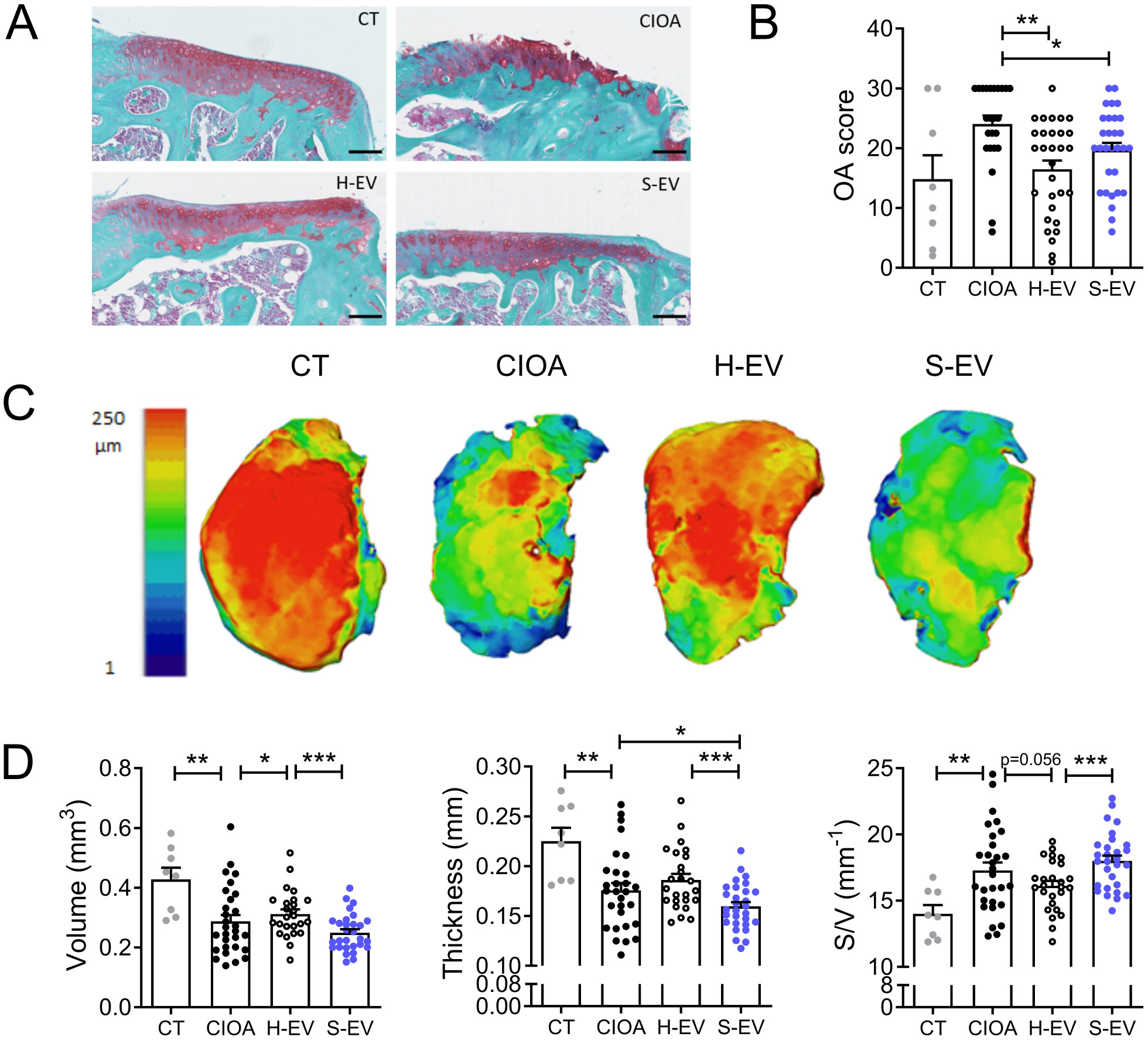
Senescent EVs fail to inhibit cartilage degradation in the collagenase-induced OA model. (A) Representative histological sections of tibias from control mice (CT), collagenase-treated mice (CIOA) and CIOA mice that received 250 ng EVs from healthy or senescent ASCs (H-EV or S-EV). Safranin O-Fast green staining. (B) Average OA score from histological sections of the different groups of mice. (C) Representative 3D reconstructed images of articular cartilage after confocal laser scanning microscopy analysis. (D) Histomorphometric parameters (volume, thickness, surface/volume) of 3D images of articular cartilage shown in (C). Data are shown as mean ± SEM (n = 8–30). Statistical analysis used the Mann-Whitney test for comparing samples pairwise with *p < 0.05, **p < 0.01, ***p < 0.001
