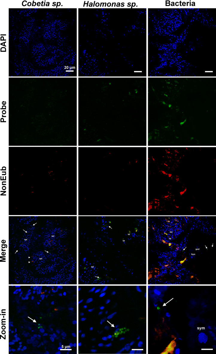
Fig 3.
Fluorescence in situ hybridization images show the specific bacterial symbionts associated with tissue samples of P. damicornis of the experimental groups treated only with saline solution (placebo) and subjected to heat treatment on the 26th day of the experiment (T2). The first column shows samples hybridized with a probe specifically designed for Cobetia sp. (in green), the middle column shows samples hybridized with a probe specifically designed for Halomonas sp. (in green), and the last column shows samples hybridized with a general Eub-338 probe (in green). All hybridizations were performed with a NonEub-338 probe (in red) to account for unspecific staining, and all sections were also stained with DAPI (in blue). Sym = Symbiodiniaceae cells; epi = epidermis; gast = gastrodermis; sp = spyrocyst; arrowhead = granular gland cell; arrow = bacteria. Scale bars in first four lines = 20 µm, scale bars at last line = 5 µm.

