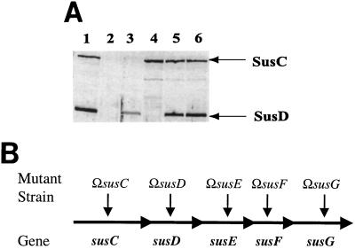
FIG. 1.
(A) Immunoblot showing SusD expression from a multicopy plasmid. Approximately 50 μg of protein was loaded in each lane. All membrane fractions were obtained from cells grown on defined medium with maltose as the sole carbohydrate source. Lanes: 1, membrane fraction from B. thetaiotaomicron 5482; 2, membrane fraction from B. thetaiotaomicron ΩsusC; 3, membrane fraction from B. thetaiotaomicron ΩsusC(pSDC27); 4, membrane fraction from B. thetaiotaomicron ΩsusD; 5, membrane fraction from B. thetaiotaomicron ΩsusD(pSDC27); 6, membrane fraction from B. thetaiotaomicron ΩsusE. This and all other immunoblots shown in this paper were scanned using an Epson Perfection 636U scanner and incorporated into a figure using both Adobe Photoshop 5.5 and Adobe Illustrator 8.0. (B) The Sus operon, showing polar insertional disruption mutations used in these studies.

