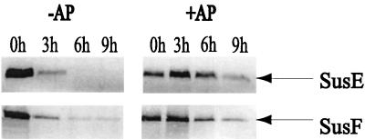
FIG. 4.
Immunoblots showing proteolytic sensitivity and protection from proteolysis by amylopectin of SusE and SusF in wild-type cells. Approximately 100 μg of protein from cell extracts was loaded in each lane. The immunoblots are labeled above according to whether amylopectin was added to the proteinase K treatment. +AP, addition of amylopectin; −AP, no amylopectin was added. The lanes are labeled according to the time that had elapsed after addition of proteinase K. In all cases, the amount of the periplasmic protein SusA was the same at all stages of digestion, and no breakdown products were detected (not shown).

