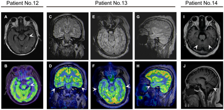
Figure 4.
The neuroimaging of patients with anti-DPPX encephalitis. (A,B) Patient 12 with cognitive impairment exhibited atrophy of the left hippocampus on magnetic resonance imaging (MRI) and decreased 18F-fluorodeoxyglucose (FDG) uptake in the left hippocampus on positron emission tomography (PET)/MRI. Patient 13 with cognitive impairment exhibited decreased 18F-FDG uptake in the bilateral temporal lobes on PET/MRI in (C,D) coronal (E,F) axial, and (G,H) sagittal views. (I,J) Patient 14 with ataxia showed cerebellar atrophy on MRI.

