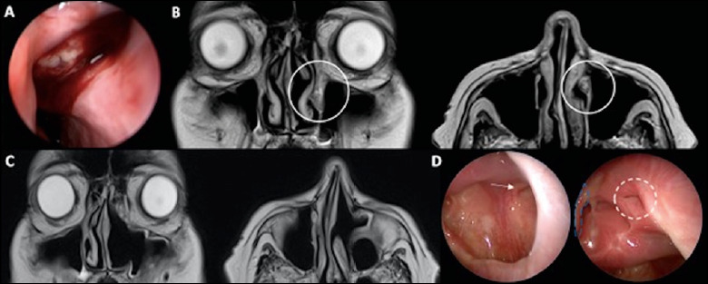
Figure 1.

A) endoscopic appearance of the lesion during first examination; B) MRI showing a polypoid mass (10 x 7 mm in axial dimension) arising from the distal nasolacrimal duct, mildly hypo-intense on T1 and T2-weighted images, and avidly enhancing on post-contrast images with hypo-intense borders; C) follow-up MRI at 6 months after surgery showing no evidence of recurrence; D) endoscopic follow-up at 6 months after surgery showing no recurrence with residual nasolacrimal duct patency (white arrow: infraorbital nerve; white ring: opening of residual nasolacrimal duct, blue line: middle turbinate residue).
