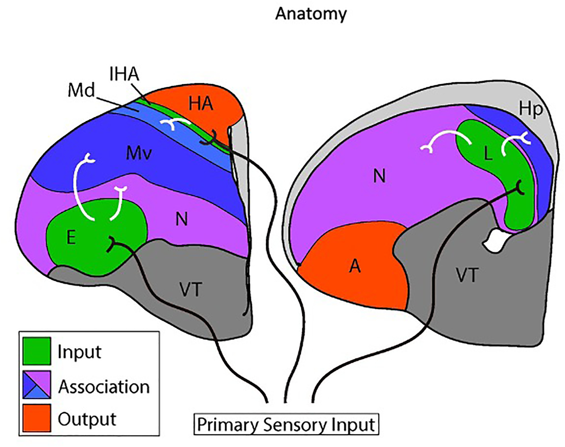
Figure 1. Connectional anatomy of the avian dorsal telencephalon.

Two coronal cross sections through the chick telencephalon are shown at intermediate (left) and posterior (right) levels. Medial is to the right and dorsal is at the top. Only the left hemisphere is shown. The two main divisions of the avian dorsal telencephalon are the Wulst and the dorsal ventricular ridge (DVR). The Wulst includes the hyperpallium apicale (HA), the interstitial nucleus of the hyperpallium apicale (IHA), and the dorsal mesopallium (Md). The DVR comprises the ventral mesopallium (Mv), the nidopallium (N), and the arcopallium (A). The hippocampus (Hp) is a medial dorsal telencephalon structure likely homologous to the mammalian hippocampus. The ventral telencephalon (VT) is indicated.
Primary input cell populations are shaded in green and output populations are red. Association territories are shaded in blue or purple. Axons originating in the dorsal thalamus (black axons) transmit visual information to the entopallium (E), auditory information to Field L (L), and visual and somatosensory information to the IHA. The Md, Mv, and nidopallium receive secondary sensory information from primary input nuclei, represented by white axons. The association territories interconnect extensively with each other and with output populations (full circuitry not shown).
