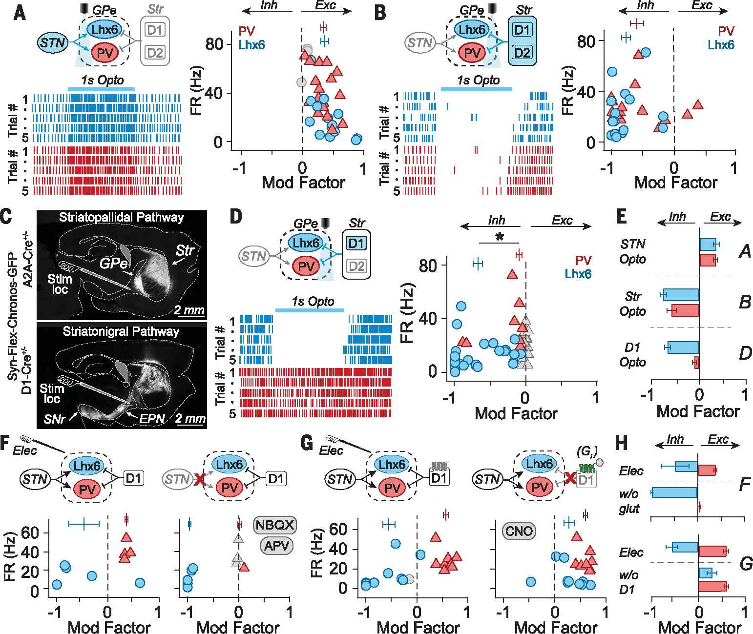
Fig. 3. Population-specific neuromodulation in the GPe is driven by convergent excitation from the STN and inhibition from D1-SPNs.

(A) (Left) Schematic and rasters from two representative GPe neurons during optogenetic stimulation of STN fibers (100 Hz for 1 s). (Right) Modulation factors (MFs) for individual neurons (symbols) and populations (vertical bars), showing that PV-GPe and Lhx6-GPe neurons are similarly excited (MFLhx6 = 0.36 ± 0.27, MFPV = 0.34 ± 0.17; MWU P = 0.976; 17 pairs of neurons; three mice). (B) (Left) Schematic and rasters from two neurons during striatal fiber stimulation (100 Hz for 1 s). (Right) MFs show that both PV-GPe and Lhx6-GPe neurons are similarly inhibited by striatal fiber stimulation (MFLhx6 = −0.77 ± 0.29, MFPV: −0.59 ± 0.41; MWU P = 0.25; 16 pairs; two mice). (C) (Top) Fluorescent image of striatopallidal pathway. (Bottom) Fluorescent image of striatonigral pathway. Typical placement of the stimulating electrode is shown for reference. (D) (Left) Schematic and rasters from two neurons during D1-SPN striatal fiber stimulation. (Right) MFs show that Lhx6-GPe neurons are preferentially inhibited (MFLhx6 = −0.68 ± 0.34; MFPV = −0.1 ± 0.23; MWU *P < 0.00001; 27 pairs; four mice). (E) Summary of MFLhx6 (blue) and MFPV (red) for experiments A to D; error bars: SEM. (F) MFs in response to electrical stimulation before (left) and after (right) application of 10 mM NBQX/50 mM APV. Excitation of PV-GPe neurons was blocked (MFCtrl: 0.36 ± 0.05, MFNBQX/APV: 0.01 ± 0.07, paired t test, P = 0.0001), but not Lhx6-GPe inhibition (MFCtrl: −0.47 ± 0.66, MFNBQX/APV: −0.96 ± 0.05, paired t test, P = 0.18) (5 Lhx6-GPe neurons, 4 PV-GPe neurons; two mice). (G) MFs in response to electrical stimulation before (left) and after (right) chemogenetic inhibition of D1-SPN fibers [AAV2-hsyn-DIO-hM4D(Gi)-mCherry + CNO, see methods]. Inhibition of Lhx6-GPe neurons was blocked (MFpre = −0.54 ± 0.39, MFCNO = 0.28 ± 0.33, paired t test, P = 0.003), but excitation of PV-GPe neurons was not (MFpre = 0.57 ± 0.15, MFCNO = 0.59 ± 0.13, paired t test, P = 0.7) (n = 10 Lhx6-GPe neurons, n= 8 PV-GPe neurons; n = five mice). (H) Summary of MFLhx6 (blue) and MFPV (red) for experiments F to G. Error bars: SEM.
