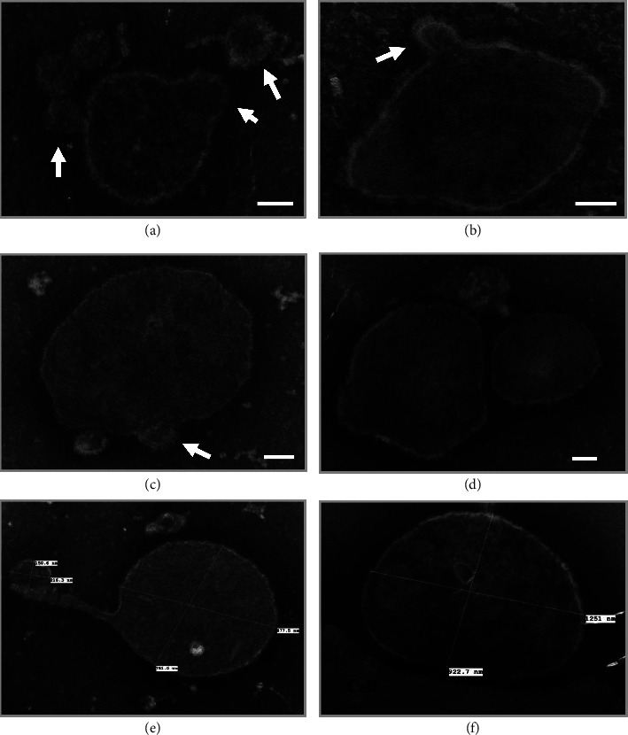
Figure 3.

Methylamine tungstate, negatively stained, whole-mount TEM analysis of mitochondria (mt) isolated from the patient's plasma. Mitochondria with different shapes and sizes, ranging from ∼400 nm (Panel (a)) to over 1 µm (Panel (f)), were identified. Some mitochondria are attached to “vesicle-like” structures protruding from the membrane (∼100 nm in diameter), pointed by white arrows in Panels (a–c). Scale bar: 100 nm.
