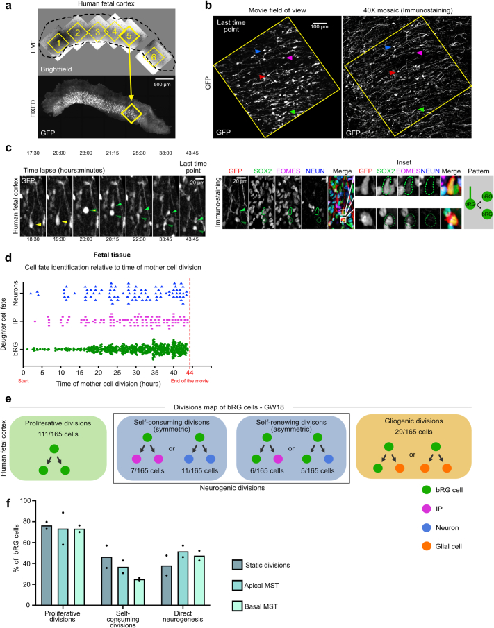
Extended Data Fig. 5. Cell fate decisions, timing and division modes in fetal tissue.
a, Automated pairing of live and fixed samples and annotation of the video fields of view on the immunostained fixed samples. b, GFP+ cell matching between the live images and the fixed images. Arrowheads indicate equivalent cells. c, Live/fixed correlative analysis of a dividing bRG cell generating two bRG daughters. d, Detection of bRG, IP or neuronal cell fate relative to the time of division of the bRG mother cell in human fetal samples at GW 14-18 (1058 cells from N = 2 fetal brains). e, Summary of all division patterns identified in bRG cells at GW 18 (N = 165 bRG cells) human frontal cortex. f, Percentage of bRG cells performing proliferative divisions, of neurogenic bRG cells performing self-consuming divisions and of neurogenic bRG cells performing direct neurogenic divisions, depending on their division mode (static, apical MST or basal MST) (N = 2 fetal brains, 415 cells). Data are presented as mean values. All images are representative examples of experiments performed in at least 2 independent fetal brains.

