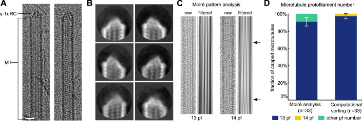
Figure 1. Cryo-EM characterisation of γ-TuRC-capped MT minus ends purified from X. laevis egg extract.
(A) Examples of γ-TuRC-capped MT minus ends identified on cryo-EM micrographs. Scale bar represents 20 nm. (B) Representative 2D classes of capped MT minus ends (mask diameter 38 nm). (C) Example raw micrograph cut-out (labelled as raw) and corresponding Moiré pattern (labelled as filtered) for a 13 protofilament (pf) and 14 protofilament MT (obtained from a non-capped MT). Arrows indicate Moiré pattern transitions characteristic of 14 protofilament MTs. (D) Fraction of Paclitaxel-stabilised, γ-TuRC-capped MTs with a specific protofilament number, as determined by Moiré pattern analysis and computational particle sorting on a subset of capped MT minus ends from n = 33 individual MTs. Error bars indicate standard deviation centred around the fraction of MTs. Source data are available online for this figure.

