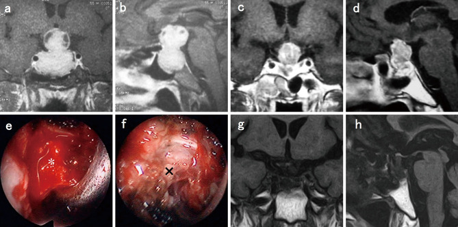
Fig. 2.
Case No. 1.
(a) Coronal T1-weighted magnetic resonance imaging (MRI) with gadolinium enhancement (Gd) revealed a pituitary tumor compressing the optic nerves. (b) Sagittal T1-weighted MRI with Gd. (c) Coronal T1-weighted MRI with Gd. (d) Sagittal T1-weighted MRI with Gd after cabergoline administration. (e) *: tumor. (f) ×: fibrous tissue after tumor resection under endoscopic view. (g) Coronal T1-weighted MRI. (h) Sagittal T1-weighted MRI after surgery.

