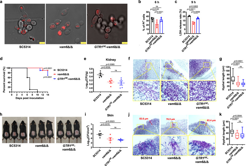
Fig. 6. Hyphal penetration forces supported by large vacuoles are required for the virulence in C. albicans.
a Imaging of vacuole membrane stained by VacuRed in C. albicans cultured in YPD medium at 30 °C for 90 min. Three biological replicates were carried out. One of the representative images are shown. Scale bars = 5 μm. b The percentage of propyl iodide (PI)-positive murine peritoneal macrophages co-incubated with C. albicans for 6 h (MOI = 1). n = 5 fields. c The LDH release rate of HUVECs co-incubated with C. albicans for 9 h (MOI = 1). n = 3 samples. d The survival percentage of mice infected with C. albicans. n = 10 mice. e The fungal burden in kidneys of mice were determined 5 days after fungal inoculation. n = 5 mice. f The kidneys of mice were taken out 5 days after fungal inoculation and made pathological sections with PAS staining. Scale bars = 100 μm. g The hyphal length in kidneys was measured by Image J. n = 20 hyphae. h The exposed back skin with fungal-infected damage on day 7. i The fungal burdens in damaged skin of mice were determined 3 days after fungal inoculation. n = 5 mice. j The pathological images of infected skin stained by PAS. The dimension of skin damage was quantified by the maximum depth of penetrated hyphae measured by Image J. Scale bars = 100 μm. k The hyphal length in damaged skin was measured by Image J. n = 20 hyphae. Data were presented as mean ± SD (b, c, e, i). Box plots indicate median (middle line), 25th, 75th percentile (box), and minimum and maximum values as well as outliers (single points) (g, k). Log-rank (Mantel-Cox) test (d), two-tailed unpaired t-test (b, c, e, g, i, k). Source data are provided as a Source Data file.

