
FIGURE 5.
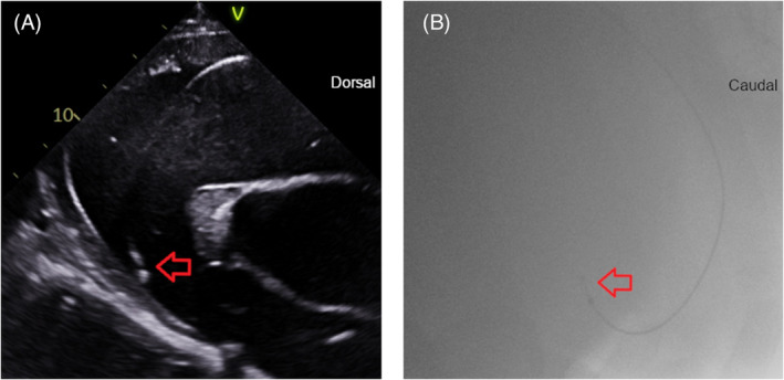
The right ventricular outflow tract lead position in selected echocardiographic and fluoroscopic images. The lead tip is denoted by the red arrow. (A) Echocardiographic right parasternal long‐axis view of the right ventricular outflow tract. Dorsal is to the right of the image. (B) Fluoroscopic right lateral image. Caudal is to the right of the image.