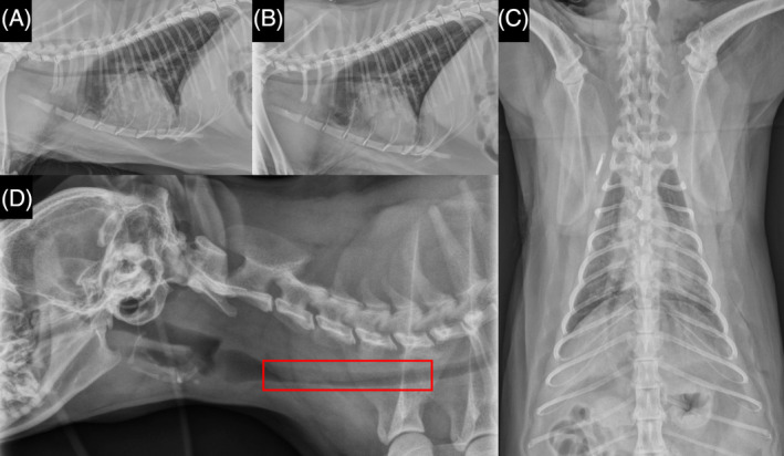
FIGURE 1.

Day 1 thoracic radiograph imaging of cat patient. Left lateral (A) and right lateral (B) and dorsoventral (C) thoracic radiographs. Note moderate, patchy, alveolar pulmonary patterns throughout the ventral aspects of the right cranial and right middle lung lobes and cranial and caudal subsegments of the left cranial lung lobe; and mild widening of the cranial mediastinum by a soft tissue opacity without a discrete mass lesion. Right lateral cervical (D) radiographic image shows a visible dorsal tracheal membrane superimposed over most of the cervical tracheal luminal diameter (red box). The cardiac silhouette, pulmonary vasculature, nasopharynx, larynx, hyoid apparatus, musculoskeletal structures, and cranial portion of the abdomen were unremarkable.
