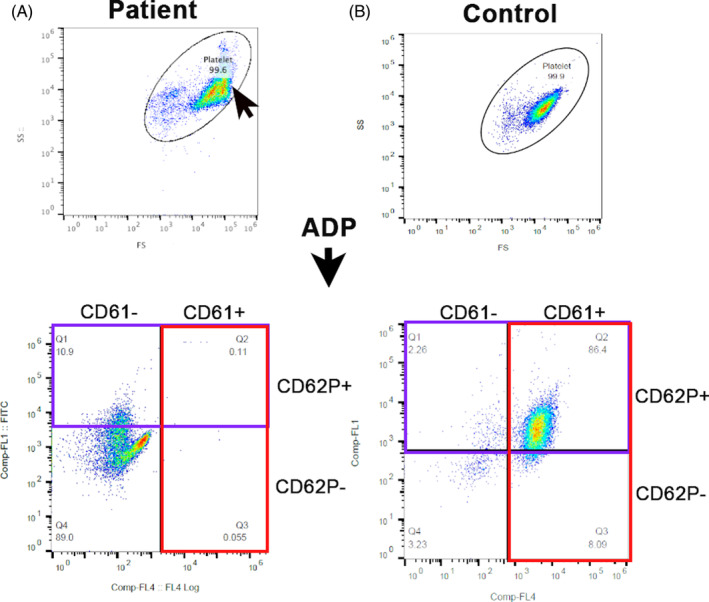
FIGURE 4.

Representative scatter dot plots of platelet flow cytometry in the patient and a control cat. Platelet gate (oval) was established using 3 and 5 μm calibration beads. (A) Note the shift in forward‐scatter (FS) in the patient (arrow) indicating an increase in platelet size compared to the control cat. Platelets were activated with ADP and labeled with fluorophore‐conjugated antibodies to αIIbβ3 (CD61; red box) and P‐selectin (CD62P, purple box). (A) A complete absence of CD61 expression (red box) was noted in the patient compared to the control cat. (B) Most platelets (86.4%) in the control cat had externalized P‐selectin (CD62+) following exposure to ADP. (A) Only a portion (10.9%) of platelets in the affected cat had externalized P‐selectin upon ADP activation. ADP, adenosine diphosphate; FS, forward‐scatter; SS, short‐scatter.
