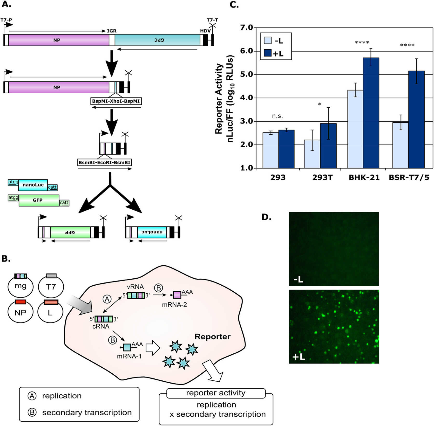
Fig. 1. Establishment of Junín virus (JUNV) minigenome systems.

(A) Schematic diagram of JUNV minigenome plasmid construction. Minigenomes were based on the S-segment of the JUNV (strain Romero) cloned in cRNA orientation into the pAmp expression vector, which contains a T7 promoter, as well as a Hepatitis Delta Virus (HDV) ribozyme and T7 terminator elements, as indicated. The open reading frames encoding the NP and GP were then excised leaving a unique cloning cassette in each location. Subsequently, either NanoLuciferase (nLuc) or green-fluorescent protein (GFP) was inserted in place of the GPC gene to generate the final minigenome constructs. (B) Schematic diagram of the JUNV minigenome system. Transfection of the minigenomes described in (A), along with pCAGGS constructs encoding the T7 polymerase, JUNV nucleoprotein (NP) and polymerase (L), leads to transcription of these three proteins by RNA polymerase II and subsequent translation. T7 then directs transcription of the minigenome RNA, which is autocatalytically processed by HDV ribozyme cleavage to generate a construct containing authentic JUNV leader and trailer sequences. This genome analogue can then be encapsidated by NP and transcribed and replicated by L. The transcribed minigenome mRNA from the GPC gene encodes an assayable reporter protein, in this case either nLuc or GFP, and the production of this reporter reflects the cumulative viral RNA synthesis (i.e. transcription and replication) taking place in these cells. (C) JUNV minigenome assay for detection of NanoLuciferase expression. The indicated cell lines were transfected as described in (B) with a monocistronic JUNV minigenome encoding nLuc as well as support plasmids, either with or without JUNV L. A control plasmid pCAGGS-Firefly (FF) was also transfected. Cells were harvested 48 h later and measured for both nLuc (viral RNA synthesis) and FF (host cell RNA synthesis) activity. The means and standard deviations of normalized reporter levels (nLuc/FF) are shown and represent data from three independent experiments. The results of a one-way ANOVA analysis to compare sample pairs with and without L are indicated. (D) JUNV minigenome assay for detection of GFP expression. Cells were transfected as described in (C) but using a GFP-expressing minigenome and omitting the pCAGGS-FF control plasmid. Samples were prepared either without (top panel) or with (bottom panel) JUNV L and fluorescence was examined after 48 h.
