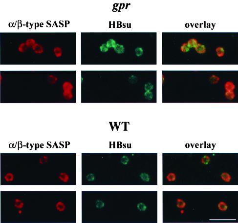
FIG. 4.
Localization of α/β-type SASP and the HBsu homolog in germinated B. megaterium spores by immunofluorescence microscopy. Wild-type (WT) and gpr (PS1551) spores of B. megaterium were germinated for 2 and 10 min, respectively, fixed, treated, immunostained for both α/β-type SASP and HBsu, and examined by fluorescence microscopy as described in Materials and Methods. The panels labeled “α/β-type SASP” show the Cy3 images, the panels labeled “HBsu” show the FITC images, and the panels labeled “overlay” show the merged images from the Cy3 and FITC images. The scale bar is 5 μm.

