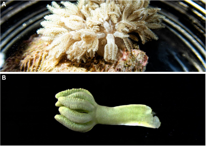
Figure 5:
Photos of Xenia umbellata samples being processed under a dissecting microscope for DNA analysis. A: A live sample from Site 1 in a glass container submerged in ocean water. B: A close-up picture of one polyp submerged in 95% ethanol and observed under the microscope. Photos by Daniel A. Toledo-Rodriguez.

