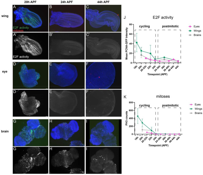
Figure 1. The timing of cell cycle exit in the Drosophila wing, eye and brain are similar.
Wings (A-C), eyes (D-F) and brains (G-I) were dissected from staged pupa and stained for mitoses (anti-Phospho Histone H3, PH3) and E2F transcriptional activity (anti-GFP for PCNA-GFP reporter) at the timepoints indicated. Animals were collected as white pre-pupa (0h APF) and incubated at 25°C to the indicated timepoints. Yellow arrowheads indicate 4 of the 8 mushroom body neuroblasts that continue to cycle until 96h APF (Siegrist et al., 2010). J-K Quantifications of E2F transcriptional activity (normalized PCNA-GFP fluorescence) and PH3 across tissues at the indicated timepoints.

