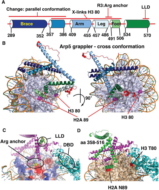
Figure 5. The LLD and arginine anchor (R3) are spatially separated in the grappler domain of Arp5.
(A) Schematic shows the alpha helices and loops that compromise the grappler domain of Arp5 based on the closed conformation. Names for different helices are shown along with the positions of the arginine anchor (R3) and LLD region as well as the region crosslinked to H2A 89 and H3 80. (B) The structure of the crossed conformation of the grappler domain of Arp5 shown and the color coding corresponds to that in (A). The location of residue 80 of H3 and 89 of H2a are highlighted. (C) The location of the arginine anchor, DNA binding domain (DBD) and the LLD region are indicated. (D) The same orientation of the grappler domain in (C) is shown with the region crosslinked to residue 80 of H3 highlighted in green and the rest of the grappler domain in purple and the actin-like portion of Arp5 in cyan.

