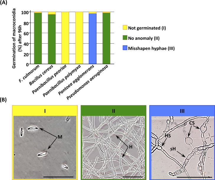
Fig 9. F. culmorum macroconidia germination and related cell structures after 96 h of exposure to rhizobacteria’s intracellular metabolites.
(A) Rate of F. culmorum’s macroconidia germinated after exposure of intracellular metabolites of the five selected rhizobacteria. (B) Example of the microscopic morphologies of macroconidia, mycelia, and chlamydospores of F. culmorum: I, not germinated; II, no anomaly; III, misshapen hyphae. M, macroconidia; H, normal hyphae; sH, septate hyphae; HS, hyphal swelling; CS, chlamydospores. Scale bars = 100 µm.

