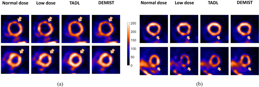
Fig. 5.
Four representative tests cases derived from four different patients, qualitatively showing the performance of TADL method and proposed DEMIST method. The short-axis slice containing the defect centroid is shown in all four cases. For all cases, the low-dose level was set to 12.5%. In (a) and (b), defects were in anterior and inferior wall, respectively. For all four cases, the defects had an extent of 30° and severity of 25%. First, we note that the background appears less noisy compared to low-dose images with both TADL and DEMIST. The defect tends to become less detectable with the TADL (no task-specific loss term). Further, the defect was visually clearer with the proposed DEMIST method.

