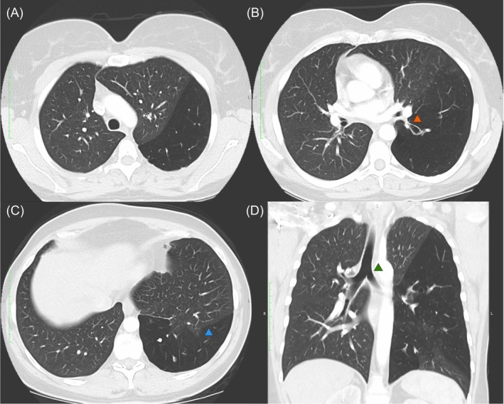
FIGURE 2.

Sagittal and coronal computed tomography images (clockwise from top left; A–D) showing left lower lobe hyperlucency and hypovascularity. (A) demonstrates hyperlucency and hypovascularity of the left lower lobe. (B) demonstrates stenosis at the origin of the left lower lobe bronchus with distal ballooning (orange arrow). (C) demonstrates an incomplete oblique fissure and subsequent parenchymal intrusion into the left lower lobe (blue arrow). In SJMS the volume of the affected lung is normal or, more commonly, reduced. It is seldom if ever increased. In this case collateral air drift through the incomplete fissure is the likely cause of the hyperinflation and subsequent mass effect. (D) demonstrates the degree of mediastinal shift with rightward tracheal deviation (green arrow). Hyperinflation is not typically a feature of SJMS and is often more suggestive of congenital lobar emphysema. If hyperinflation is present, this suggests presence of collateral air flow either from adjacent unaffected lung, or across an incomplete fissure as seen in this case.
