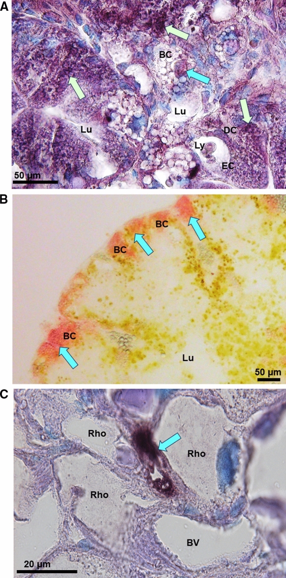
Fig. 7.

Light microphotographs of midgut gland tissue sections of Helix pomatia. A Midgut gland cells of tubular epithelia showing the Lumen (Lu) surrounded by Basophilic Cells (BC), Digestive Cells (DC) and Excretory Cells (EC) with CdMT mRNA expression signals (dark violet spots) indicated by light green arrows, and a condensed cell body in programmed cell death within a basophilic cell (light blue arrow), B Midgut gland tubulus with central Lumen (Lu) and peripheral Basophilic Cells (BC) with Zn visualized by Dithizone reactions (blue arrows and red color). C Midgut gland section showing a group of Rhogocytes (Rho) delimiting a Blood Vessel (BV), with CuMT mRNA expression signals (blue arrow, dark violet spot)
