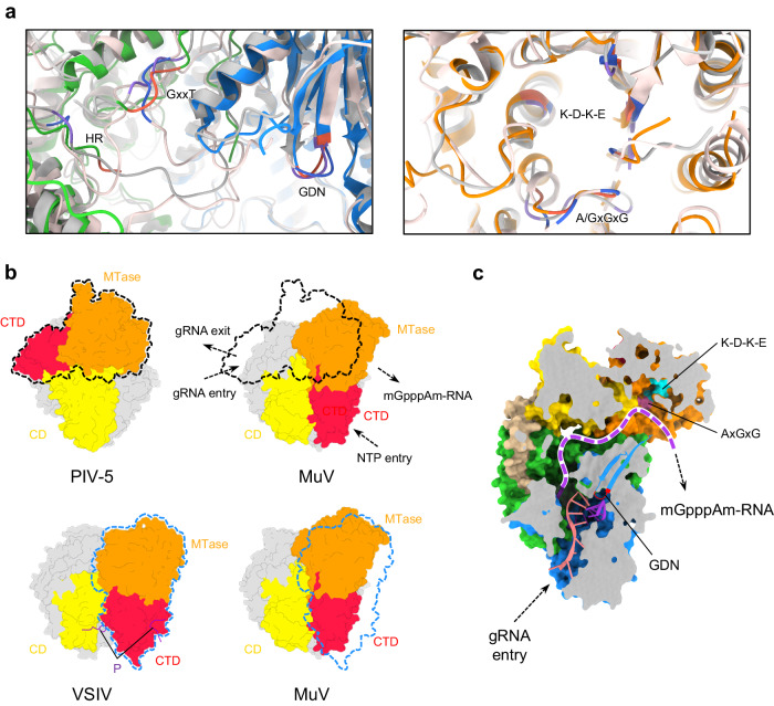
Fig. 2. MuV Lintegral–P as a favorable transcription state.
a Comparison of critical motifs among MuV, PIV-5, and VSIV L. Motifs (GDN, GxxT, HR, K-D-K-E, and A/GxGxG) of MuV, PIV-5, and VSIV are colored in tomato, medium purple, and royal blue, respectively; the other parts of PIV-5 and VSIV are colored in silver and misty rose, respectively. b Comparison of CD-MTase-CTD spatial organizations among MuV, PIV-5, and VSIV L. RdRp and PRNTase of all three structures are aligned and colored in light gray. The P fragment of VSIV is colored in purple. The outlines of PIV-5 and VSIV MTase-CTD are depicted in black and blue dashed lines around MuV maps, respectively. c Continuous RNA tunnel of MuV Lintegral–P. Superposed nucleotides are from the crystal structure of the reovirus λ3 polymerase initiation complex (PDB ID 1N1H). The purple dashed curve represents the potential elongation path for the transcribed mRNA.

