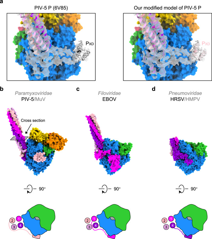
Fig. 4. Diverse origins of L-binding PXD in nsNSVs.
a The atomic model of PIV-5 P (PDB ID 6V85) docked into the PIV-5 P density (EMD-21095) (Left) and the modified atomic model of PIV-5 P based on the atomic model of MuV P (Right). b The side view of our newly built atomic model of PIV-5 L–P complex. The cartoon demonstrates the top view of the L–P interface. Four circles represent the cross-sections of the P tetramer. The rounded rectangle represents the PXD. c The side view of the atomic model of the EBOV L–P complex. The top view of the L–P interface is shown in the cartoon style. d The side view of the atomic model of the HRSV L–P complex. The top view of the L–P interface is shown in the cartoon style.

