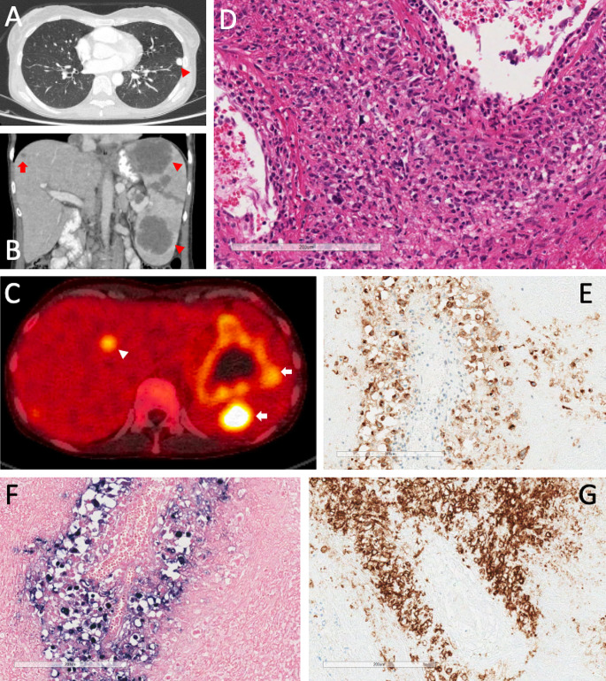
Fig. 1.
(a) CT scan through chest showing pulmonary granulomatous lesion (arrow head). (b) CT through abdomen showing lesions affecting the spleen (arrow heads) and liver (arrow). (c) PET scan though abdomen showing avid FDG lesions in the spleen (arrows) and liver (arrow head) in both organs. (d) Splenic biopsy (haematoxylin/eosin) showing large atypical lymphoid cells with an angiocentric growth. (e) Splenic biopsy CD30 stain highlighting the neoplastic B cells. (f) Splenic biopsy EBER stain. (g) Splenic biopsy CD20 stain showing neoplastic B cells with an angiocentric arrangement

