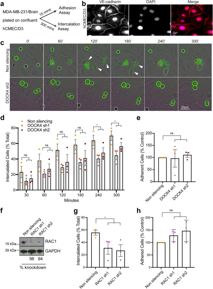
Fig. 2. Knockdown of DOCK4 or RAC1 blocks breast cancer cell intercalation into brain endothelial cells.
a Schematic depicts parallel in vitro adhesion and intercalation assays following seeding of MDA-MB-231/Brain cells onto confluent human brain endothelial cells (hCMEC/D3). Adhesion was determined 15 min and intercalation 5 h post-seeding. b Immunofluorescence images of confluent hCMEC/D3 monolayer prior to seeding MDA-MB-231/Brain cells. Adherens junctions (VE-cadherin) were visualised. Scale bar = 20 µm. c Still phase images from timelapse movies (see Supplementary Movie 2) showing MDA-MB-231/Brain cells (green) transduced with either control (Non silencing) or DOCK4 (sh2) shRNAs seeded onto confluent hCMEC/D3. White arrowheads indicate elongated cells prior intercalation. Scale bar = 20 µm. d Graph shows quantification of MDA-MB-231/Brain cells intercalated into hCMEC/D3 (% total) from timelapse movies. e Graph shows adhesion of MDA-MB-231/Brain cells 15 min post-seeding onto confluent hCMEC/D3. Data expressed as percentage of control (non silencing). f Immunoblot analysis of MDA-MB-231/Brain cells following RAC1 stable depletion (RAC1 sh1, RAC1 sh2). g Graph shows quantification of MDA-MB-231/Brain cells intercalated into hCMEC/D3 (% total) from timelapse movies at the timepoint when ≥50% of control (non silencing) cells intercalated into hCMEC/D3. h Graph shows adhesion of MDA-MB-231/Brain 15 min post-seeding onto confluent hCMEC/D3. Data are expressed as the percentage of control (non silencing). d, e, g, h Error bars represent SEM from N = 3 independent experiments in which ≥100 cells were analysed from ≥9 movies per condition in 3 technical replicates per experiment. *P < 0.05 by two-tailed Student’s t test.

