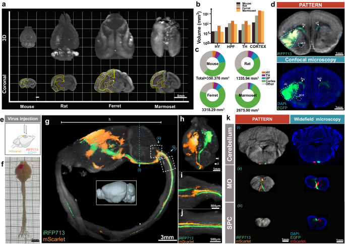
Fig. 3. PATTERN-based whole-brain optical imaging.
a Representative 3D whole-brain imaging of mouse, rat, ferret, and marmoset via PATTERN. b, c Geometric analysis of the representative mouse, rat, ferret, or marmoset brain according to data acquired by PATTERN. HY Hypothalamus, HPF Hippocampus Formation, TH Thalamus. d Validation of PATTERN-acquired fluorescence signal. e Dual-color labeling strategy for visualizing projections from the motor cortex to the spinal cord. Partially created with BioRender.com released under a Creative Commons Attribution-NonCommercial-NoDerivs 4.0 International license https://creativecommons.org/licenses/by-nc-nd/4.0/deed.en. f Bright field images of entire central nervous system. 3D-view images by PATTERN of the entire central nervous system (g), brain only (h), medulla oblongata (i), and spinal cord (j). k Coronal section via PATTERN (left column) and widefield microscopy (right column), medulla oblongata (MO), and spinal cord (SPC). Positions were labeled by roman numerals corresponding to (g).

