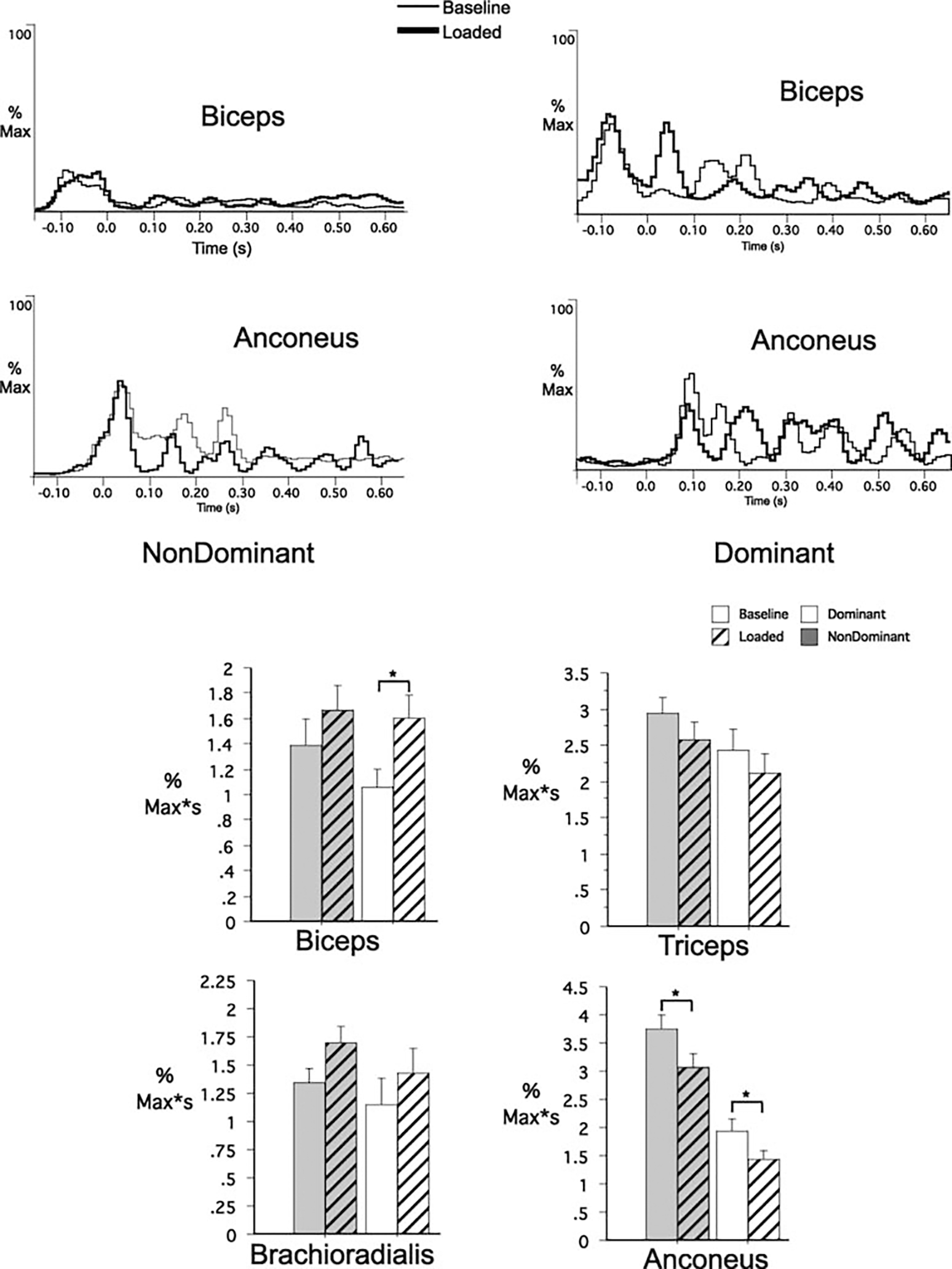
Fig. 3.

EMG recordings (individual trials) for biceps brachii and anconeus for the nondominant (left) and the dominant (right) arm under naïve performances. Data were synchronized to elbow peak acceleration. Data were normalized to the maximum muscle activity. Bar graphs: EMG impulse comparisons for dominant and nondominant arm groups for all recorded muscles. *Results from statistical analysis were significant
