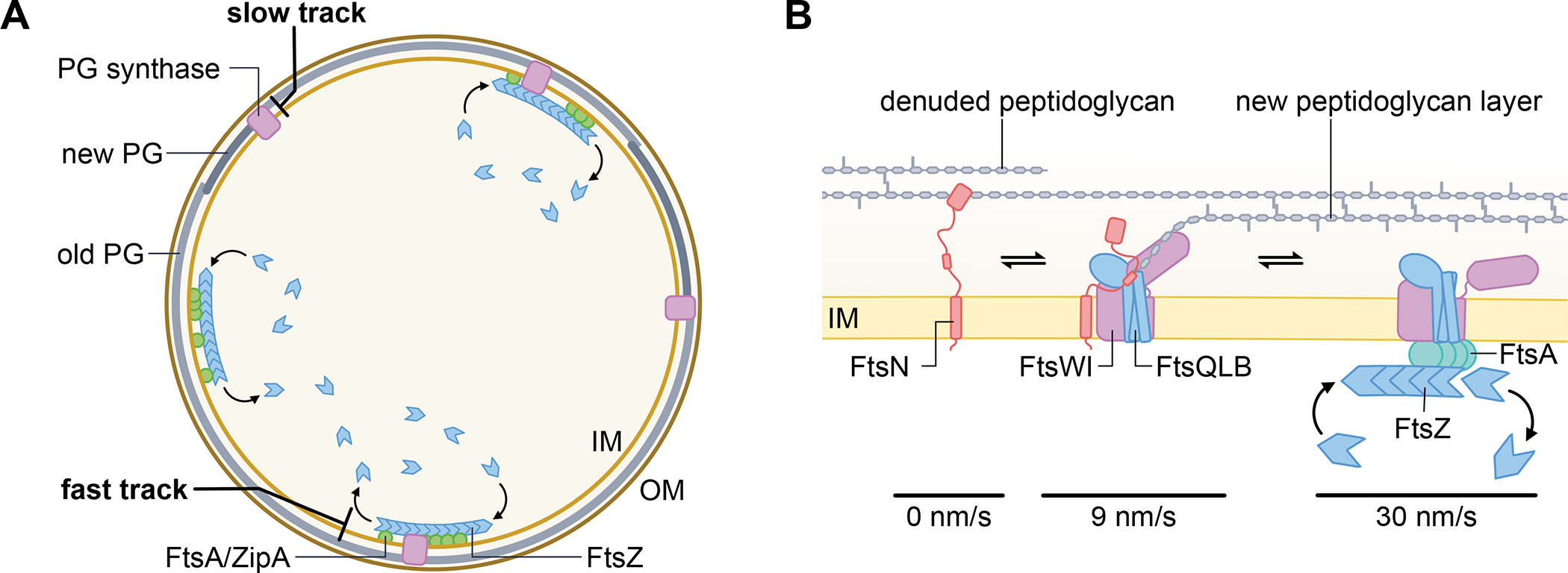
Fig. 4. Divisome proteins build the septum in two tracks.

(A) Cross section of a dividing E. coli cell depicting several processive, FtsZ treadmilling complexes that move rapidly along the cytoplasmic face of the inner membrane to organize septum synthesis, in coordination with several independently moving, slower track of proteins that actively engage in septal peptidoglycan (PG) synthesis in the periplasm. (B) Detail of complexes comprising the different speed modes during septum formation. A subset of FtsN proteins are transiently stationary, possibly because they are bound to denuded septal PG resulting from amidase activity. Another subset of FtsN proteins is on the slow track (~9 nm/s) when in complex with FtsQLB and FtsWI as they synthesize septal PG, activated by FtsN. On the right is a putative fast moving (~30 nm/s) complex containing FtsZ and FtsA that spatially guides FtsQLB and FtsWI so the latter are properly placed to be handed off to the slow track for septum synthesis.
