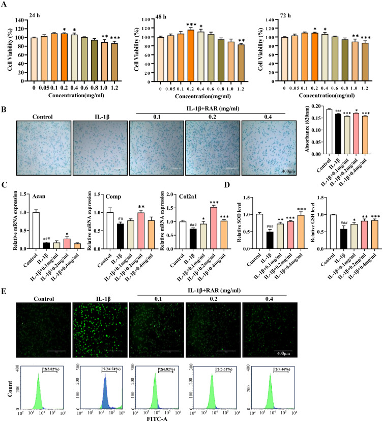
Figure 4.
RAR promotes chondrocyte anabolism and inhibits oxidative stress in vitro. (A) CCK-8 was used to measure cell proliferation following 24, 48, and 72 h of RAR treatment. (B) Alcian blue staining was used to detect acidic polysaccharides at various RAR concentrations. (C) Acan, Comp, and Col2a1 relative mRNA expression in IL-1β-induced primary chondrocytes treated with RAR. (D) Bar graph of SOD and GSH changes. (E) ROS generation in chondrocytes treated with various RAR concentrations was measured using the ROS probe DCFH-DA and quantified using a flow cytometer. Control vs IL-1β ##P < 0.01, ###P < 0.001; IL-1β vs RAR *P <0.05, ** P <0.01, *** P<0.001.

