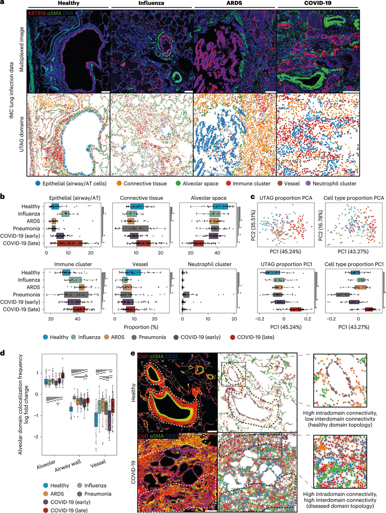
Fig. 3 |. Microanatomical domains discovered by UTAG across data types and disease states.

a, Discovery of microanatomical domains in IMC images of lung from patients of various pathologies. The top row illustrates the intensity of three selected channels and the bottom row displays the UTAG domains. Scale bars, 200 μm. b, Univariate analysis of microanatomical domain composition across lung infection disease. Microanatomical domain composition was percent normalized per slide. c, PCA for joint analysis of domain (left) or cell type (right) composition per image. The top two plots visualize the position of images in the first two principal components. The bottom two plots show the distribution of the first principal component aggregated by disease group. d, Log odds of domain colocalization frequencies across diseases in alveolar domains. Log odds indicates observed frequency over expected, as estimated empirically by random permutation. Positive values indicate high intradomain (alveolar–alveolar) colocalization compared to random mixtures and negative indicates low interdomain colocalization. **P < 0.01, *P < 0.05, two-sided Mann-Whitney U-test after Benjamini-Hochberg adjustment. e, IMC images of healthy and COVID-19 infected lung tissue. The image of healthy lung tissue shows highly compartmentalized domains, particularly in the vasculature, while the image of the diseased lung shows loss of compartmentalization. Scale bars, 200 μm. For b and c, n = 239 highly multiplexed IMC lung images from 27 deceased patients due to lung infection. *P < 0.05, two-sided Mann-Whitney U-test after Benjamini-Hochberg adjustment. Data in boxplots are presented by minimum, 25th percentile, median, 75th percentile and maximum. Values outside of 1.5 times interquartile range are classified as outliers and are denoted as fliers. ARDS, acute respiratory distress syndrome.
