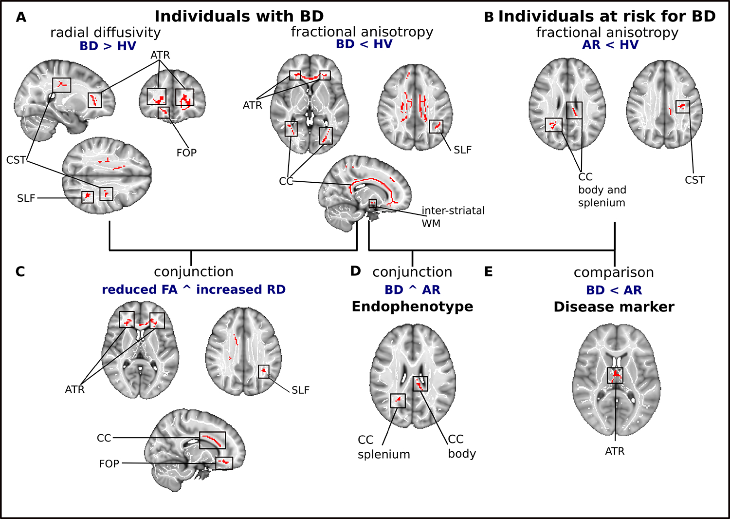
Figure 1. Depiction of differences and communalities in white matter microstructure between individuals with and at risk for BD, and healthy volunteers.

(A) Clusters of reduced fractional anisotropy and increased radial diffusivity in individuals with BD compared to healthy volunteers. (B) Clusters of reduced fractional anisotropy in individuals at risk for BD. (C) Results of multimodal analysis depicting clusters with both reduced fractional anisotropy and increased radial diffusivity in individuals with BD. (D) Regions of reduced fractional anisotropy in individuals with or at risk for BD, compared to HV. (E) WM tracts with more pronounced FA reductions in BD than AR. Significant clusters are overlaid on the MNI template and a standardized fractional anisotropy skeleton.
Abbreviations: AR, individuals at risk for bipolar disorder; ATR, anterior thalamic radiation; BD, bipolar disorder; CC, corpus callosum; CST, corticospinal tract; FOP, fronto-orbito polar tract; PTR, posterior thalamic radiation; SLF, superior longitudinal fasciculus; WM, white matter
